Back Bones Labeled - Shoulder Bones Labeled Stock Photo Picture And Royalty Free Image Image 43229690 - Throughout the spine, intervertebral discs made of.
Back Bones Labeled - Shoulder Bones Labeled Stock Photo Picture And Royalty Free Image Image 43229690 - Throughout the spine, intervertebral discs made of.. The vertebrae, which stack like spools of thread, support the back and protect the spinal cord. Learn the major cranial bone names and anatomy of the skull using this mnemonic and labeled diagram. The part of the nerve that emerges out of the spine is called the nerve root. The vertebral column, also known as the backbone or spine, is part of the axial skeleton. There are 33 vertebrae in total;
The 26 bones of spine are called vertebrae. The spine is composed of 33 bones called vertebrae, which stack together to form the spinal canal. The spine anatomy is a complex structure. The 5 sections of the spine from the top to bottom are: The vertebral column, also known as the backbone or spine, is part of the axial skeleton.
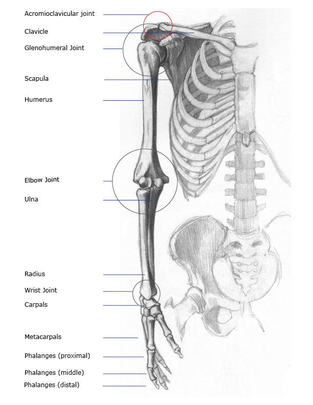
Arm Bones Joints Front Anterior And Back Posterior Anatomy Views from www.healthpages.org By adulthood, it is a large, triangular bone, that forms the base of the spinal column where it connects to the pelvic bones. The first 5 bones of spine are known as the cervical vertebrae, the next 12 bones are known as the thoracic vertebrae followed by 5 lumbar vertebrae and then one fused sacral and a coccyx at the last. See human back anatomy stock video clips. Conditions of the lumbar spine Facet joints connect each vertebra, with fluid supporting. In the back and elsewhere in the body, tendons attach muscles to bones. The bones of the back, together, make up the vertebral column.the vertebral column is made up of 5 sections: The part of the nerve that emerges out of the spine is called the nerve root.
The column can be divided into five different regions, with each region characterised by a different vertebral structure.
The first 5 bones of spine are known as the cervical vertebrae, the next 12 bones are known as the thoracic vertebrae followed by 5 lumbar vertebrae and then one fused sacral and a coccyx at the last. This protects the spinal cord inside. The spine anatomy is a complex structure. Your spinal cord is protected by the vertebral column (spinal column or backbone). This vertebra supports the skull. Foot bone anatomy x ray 12 photos of the foot bone anatomy x ray foot bone anatomy x ray, bone, foot bone anatomy x ray. What are the 5 sections of the spine? Vertebrae are the structural constituents of the spine. By adulthood, it is a large, triangular bone, that forms the base of the spinal column where it connects to the pelvic bones. Learn the major cranial bone names and anatomy of the skull using this mnemonic and labeled diagram. Atlas (c1) the atlas is the first cervical vertebra and therefore abbreviated c1. This part of your anatomy is susceptible to injury, arthritis, herniated disks, pinched nerves and other problems. Below the lumbar spine is a bone called the sacrum, which makes up the back part of the pelvis.
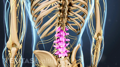
Nerves in your lower back five pairs of lumbar spinal nerves labeled l1 to l5 branch off your spinal cord and exit through small holes between the vertebrae. But, they are common in the back and can cause pain. Top free images & vectors for skull bones labeled back in png, vector, file, black and white, logo, clipart, cartoon and transparent. The sacrum is a flat, triangular bone found in the lower back and wedged between the 2 hip bones. Browse 222 lower back skeleton stock photos and images available, or start a new search to explore more stock photos and images.
Understanding Lower Back Anatomy from embed.widencdn.net Human body anatomy female female anatomy muscle shoulder blade pain anatomy back muscles bones man female anatomy body muscles in a body female anatomy muscole shoulder concept muscular sysyem. The bones of the back, together, make up the vertebral column.the vertebral column is made up of 5 sections: The cervical vertebrae, the thoracic vertebrae, the lumbar vertebrae, the sacrum and the coccyx.these sections total 33 vertebrae which function together to aid locomotion and posture as well as providing support and protection. Facet joints connect each vertebra, with fluid supporting. By adulthood, it is a large, triangular bone, that forms the base of the spinal column where it connects to the pelvic bones. Nerves in your lower back five pairs of lumbar spinal nerves labeled l1 to l5 branch off your spinal cord and exit through small holes between the vertebrae. See lumbar spine anatomy diagram stock video clips. The pelvis is composed of the two pelvic bones and the sacrum and coccyx (the pelvic bones are also known as the coxal, innominate, or hip bones) (fig.
The 5 sections of the spine from the top to bottom are:
Back, bones and human spin diseases explanation vector. The twelve thoracic vertebrae are numbered t1 to t12. See human back anatomy stock video clips. These bones work together to provide flexibility to the trunk, support the muscles of the trunk, and protect the spinal cord and spinal nerves of the back. Muscle or tendon injuries can occur anywhere in the body. The remaining small bones or ossicles below the sacrum are also fused together and called the tailbone or coccyx. The vertebral column of the lower back includes the five lumbar vertebrae, the sacrum, and the coccyx. In the back and elsewhere in the body, tendons attach muscles to bones. See sacrum (sacral region) the sacrum is connected to part of the pelvis (the iliac bones) by the sacroiliac joints. The occiput (co), also known as the occipital bone, is a flat bone that forms the back of the head. This bone is shaped like a triangle that fits between the two halves of the pelvis, connecting the spine to the lower half of the body. They help support particular bones and make them move. Foot bone anatomy x ray 12 photos of the foot bone anatomy x ray foot bone anatomy x ray, bone, foot bone anatomy x ray.
The remaining small bones or ossicles below the sacrum are also fused together and called the tailbone or coccyx. They help support particular bones and make them move. The lumbar spine is composed of five. The bones of the back, together, make up the vertebral column.the vertebral column is made up of 5 sections: Human bone jewelry 12 photos of the human bone jewelry human bone jewelry, human bone jewelry legal, human.
How To Learn The Human Bones Tips To Memorize The Skeletal Bones Anatomy Physiology Youtube from i.ytimg.com See sacrum (sacral region) the sacrum is connected to part of the pelvis (the iliac bones) by the sacroiliac joints. Learn the major cranial bone names and anatomy of the skull using this mnemonic and labeled diagram. Posterior view of the lumbar spine and pelvis. The sacrum is a flat, triangular bone found in the lower back and wedged between the 2 hip bones. The lumbar spine is composed of five. The five lumbar vertebrae are numbered l1 to l5. The range of motion in the thoracic spine is limited. Label the bones on the skeleton.
The vertebral column of the lower back includes the five lumbar vertebrae, the sacrum, and the coccyx.
There are 33 vertebrae in total; Conditions of the lumbar spine 294 label search results for back bone. The column can be divided into five different regions, with each region characterised by a different vertebral structure. Below the lumbar spine is a bone called the sacrum, which makes up the back part of the pelvis. The pelvis is composed of the two pelvic bones and the sacrum and coccyx (the pelvic bones are also known as the coxal, innominate, or hip bones) (fig. In the back and elsewhere in the body, tendons attach muscles to bones. The part of the nerve that emerges out of the spine is called the nerve root. This part of your anatomy is susceptible to injury, arthritis, herniated disks, pinched nerves and other problems. By adulthood, it is a large, triangular bone, that forms the base of the spinal column where it connects to the pelvic bones. Vertebrae separated by intervertebral discs. But, they are common in the back and can cause pain. Your healthcare provider can help ease back pain and offer.
But, they are common in the back and can cause pain back bones. See lumbar spine anatomy diagram stock video clips.


0 Komentar